
Postsynaptic cells innervate the pupillary sphincter, resulting in miosis.

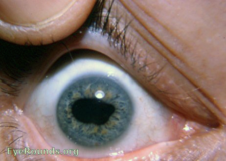
This structure connects bilaterally, but predominantly contralaterally, to the oculomotor nuclear complex at the Edinger–Westphal ( E-W ) nuclei, which issue parasympathetic fibers that travel within the third nerve (inferior division) and terminate at the ciliary ganglion ( CG ) in the orbit. Light entering one eye ( straight dark arrow, bottom right ) stimulates the retinal photoreceptors ( RET ), resulting in excitation of ganglion cells, whose axons travel within the optic nerve ( ON ), partially decussate in the chiasm ( CHI ), then leave the optic tract ( OT ) (before the lateral geniculate nucleus ( LGN )) and pass through the brachium of the superior colliculus ( SC ) before synapsing at the mesencephalic pretectal nucleus ( PTN ). Pupillary light reflex-parasympathetic pathway. The ipRGCs are most sensitive to blue light, and the preservation of circadian rhythms and the pupillary light reflexes in patients with severe photoreceptor diseases and Leber’s hereditary optic neuropathy can be explained by intact ipRGC function. Additionally, intrinsically photosensitive retinal ganglion cells (ipRGCs) containing melanopsin, a photopigment, can be activated by light without photoreceptor input. Light entering the eye causes retinal photoreceptors to hyperpolarize, in turn causing activation of retinal interneurons and ultimately the retinal ganglion cells. Normally, light directed at either eye leads to bilateral pupillary constriction, and this pupillary light reflex is mediated by a parasympathetic pathway (see Fig. The sphincter muscle wraps 360 degrees around the pupillary margin, and the dilator muscle similarly encircles the pupil but is more peripherally located. Contraction of the dilator muscle leads to pupillary enlargement (mydriasis), while sphincter muscle contraction causes pupillary constriction (miosis). The iris contains the two muscles that control the size of the pupil. Note the pupil is slightly nasal to the center of the cornea and iris.


 0 kommentar(er)
